Professional Resources
Abreakthrough in ultrasound breast imaging. ABUS is an ultrasound breast imaging technology that is less affected by a woman’s breast density – providing a clearer and more insightful image. Unlike mammography, which uses radiation, ABUS uses sound waves to create 3D images of the breast tissue.The ABUS ultrasound device is FDA and Health Canada cleared for use by doctors in combination with mammography and is most commonly used when a woman has known breast abnormality or symptom.
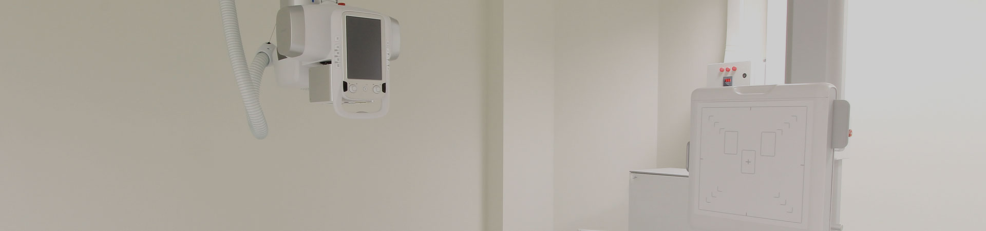
ABUS delivers uncompromised image quality. ABUS is specifically developed and designed for breast imaging. Incorporating the latest automated ultrasound technology, ABUS delivers uncompromised image quality that is increasing confidence in breast ultrasound. ABUS is used as an adjunct to mammography for screening asymptomatic women for Breast Cancer. The system works by positioning a sheer membrane gently on the breast after application of a conductive gel. It then automatically images the whole breast after which images can be rendered and reviewed in 3D. The complete field of view covers 15cm x 17cm x 5.0cm. The machine has a high center-frequency enabling high resolutions and ultra broadband characteristics for optimal contrast.
ABUS’ standardized automatic scan virtually eliminates human error – the large size probe captures images in precise anatomical detail of complex breast tissues and structures without relying solely on the skills of the operator. The standard quality of ABUS is exponentially better and more accurate than human operated ultrasound systems used elsewhere and very powerful for assessment of the entire breast.
Mammogram alone may not be enough for some of your patients. ABUS is not meant as a replacement of mammography for screening, but rather as an adjunct in patients with dense breast tissue to detect small tumors. Women with dense breasts are at increased risk for Breast Cancer. It is widely accepted that up to 40% of North American women and up to 70% of Asian women have dense breasts. ABUS is an advanced technology approved in the United States and the European Union as a consistent, accurate 3D ultrasound breast screening tool in the early detection of Breast Cancer. The detection rate of cancer lesions smaller than 1cm is increased 50% by ABUS to that of mammogram alone.
ABUS is not just for women with dense breasts. ABUS is safe and radiation free for all women at every stage in their life. ABUS can be used safely for younger, pregnant, lactating, post-operative or women who are high risk and would otherwise be adverse to receiving a radiation dose from a mammogram. ABUS is the test of choice for the assessment of women with breast implants, post-operative, or scarred breast tissue. The ABUS system can be used as frequently as needed for follow up scans.
SOMO−INSIGHT Clinical Study. A large trial, the SOMO−INSIGHT Study, is currently underway in the United States to determine whether digital mammography in combination with the Automated Breast UltraSound system or a routine screening mammogram alone is more sensitive in detecting Breast Cancer in women with greater than fifty percent dense breast tissue. Up to 20,000 patients will be enrolled in the study.
EASY Clinical Study. The EASY Clinical Study (European Asymptomatic Screening Study) is a large European clinical research study sponsored by U-Systems Inc. to evaluate the integration of ABUS into the routine practices of a high-volume hospital-based breast cancer screening program in Stockholm, Sweden. The clinical study is designed to determine whether Full Field Digital Mammography combined with ABUS can improve breast cancer detection when compared to mammography alone in women with dense breast tissue.
This study is particularly relevant to the many population based screening programs in Europe which practice a double-reading methodology for screening, where two different radiologists independently review breast imaging exams. The study protocol has accommodated the standard screening practices in Sweden and outlined a process for integrating ABUS review into an existing double-read system. Screening mammography, the mainstay for breast cancer detection, has known limitations in women with dense breast tissue and these women have a higher risk of breast cancer. A new approach is needed to improve breast cancer detection for women with increased breast density. The EASY breast cancer screening study will help determine if using this 3D ultrasound imaging technology in combination with a digital mammogram could improve the accuracy of routine screenings for women with dense breast tissue, for which mammography can be less effective.
The study intends to recruit 8,000 asymptomatic women with dense breast tissue. While the results of the primary analysis are not intended to reach statistical significance, they will yield valuable trending information about the potential impact on existing cancer detection rates a multi-modality approach combining ABUS with routine mammography may have for the group of asymptomatic women with dense breast tissue who participate in population-based service screening in Sweden.
SOMO−INSIGHT Study. SOMO−INSIGHT is a multi-center nationwide clinical research study sponsored by breast cancer screening Toronto to evaluate if the Automated Breast UltraSound (ABUS) done together with a routine screening mammogram (x-ray) is more accurate in detecting Breast Cancer in women with dense breast tissue than having a routine screening mammogram alone.
The fully automated ultrasound technology, which is used in the clinical research study, is cost effective and patient friendly. The software creates 3D-reconstructed images of the breast tissue for the radiologist to review. Using U-Systems software, the radiologist can look through hundreds of breast tissue image “slices” virtually peeling back the layers of dense tissue to visualize cancers, which may have been hidden by that tissue on the mammogram.
Breast density has also been shown to increase a women’s lifetime risk for developing Breast Cancer. About 40% of women have some dense breast tissue, and visualization of cancers in dense breast tissue with mammography is sometimes limited. The result is missed cancers or the discovery of later-stage cancers in women which may require more aggressive treatment options. A more thorough screening approach may be needed for these women and the SOMO−INSIGHT clinical research study is exploring a possible solution. According to a peer reviewed study by Boyd et al published in the New England Journal of Medicine, “Women with dense tissue in 75% or more of the breast have a risk of Breast Cancer four to six times as great as the risk among women with little or no dense tissue. While mammography remains the gold standard for Breast Cancer screening, clinical studies have shown improved early detection of Breast Cancer when ultrasound is used as an adjunct to mammography for women with dense breast tissue. The SOMO−INSIGHT Clinical Study has identified a significant number of mammographically negative Breast Cancers that would not have been detected had the participants not had an ABUS exam.
Medical Information
ABUS 3D images provide clinicians the ability to thoroughly and efficiently examine breast tissue. The format may seem unfamiliar at first. Click here to learn more about the ABUS image display and image manipulation options. The ABUS scan head assembly utilizes disposable, single-use mesh membranes to aid in acoustic coupling, compression and stabilization. Learn more by contacting GE Accessories at 1-800-558-5210.
ABUS has been shown to find small, invasive, node-negative cancers that were missed by mammography. ABUS as a supplemental exam is becoming an integral part of many practices. Click here to learn more about the clinical benefits of ABUS, including research, case studies, webinars and testimonials.
Product Monography
What is special about ABUS technology?
Technology advancements: Using proprietary technology to automate the ultrasound imaging process, the ABUS system was created specifically for breast screening. Advanced algorithms automate the imaging process to help provide remarkable image quality and reproducibility from user to user.
Reading station: Developed specifically for the high-volume, breast cancer screening environment. The advanced 3D ABUS Workstation enables fast, accurate review and archiving of patient exams, optimizing the breast ultrasound screening workflow.
How does ABUS benefit physicians?
With improved contrast resolution, the ABUS has the potential to reduce false positives and improve confidence when characterizing lesions.
With the coronal view capabilities provided by ABUS, clinicians have the ability to interrogate suspicious areas in a 3D plane. The ability to see the entire breast, not just the sample images, gives clinicians greater control over the review process, supporting more confident decisions.
IABUS Single-Use Stabilization Membrane
The ABUS disposable scan membrane provides comfort to the patient during the 3D image acquisition. The flexible, polyester, mesh fabric stabilizes the breast tissue and enables better image quality during the scan. The membrane is single-use-only which eliminates the risk of cross-contamination and the need for sterilization. Curved membrane dimensions: 229mm (L) x 213mm (W) x 19mm (D).